
When people talk about animal testing, the conversation almost always drifts to cosmetics, and for good reason; testing lipstick or shampoo on animals is now widely, and rightly, seen as unnecessary. But that debate has quietly swallowed a bigger, more relevant one for anyone in veterinary medicine: the role animal research plays in biomedical science, including the science that treats the patients on our own exam tables.
For a veterinary audience, this isn't an abstract ethics debate about human hospitals. Dogs were the model animal in the room when insulin was discovered. Dogs are still the model animal used to test pacemaker leads today. And in a loop most pet owners never hear about, dogs are also the patients who now receive hip replacements and pacemakers in general veterinary practice, treatments built on the very research tradition people associate with human medicine alone.
This piece walks through three of those technologies: what was actually learned from the animal model in each case, why it couldn't have been learned another way, how veterinary research sits inside the same oversight framework as human biomedical research, and what that means for how we talk to clients about the medicine we practice.
1. Cardiac Pacemakers: Tested in Dogs, Now Implanted in Dogs
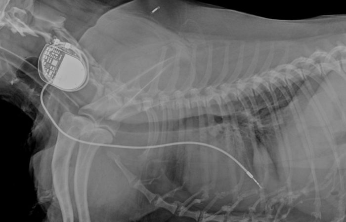
Image credit: Veterinary Thought Exchange (VTX CPD). Available at: https://vtx-cpd.com
Built on canine models
Modern pacemaker technology traces back to canine experiments. Early work showing that a failing heart's rhythm could be regulated with external electrical stimulation used dogs as the model, before later testing moved to species like the guinea pig and pig as the technology miniaturized.
That lineage is still active. A standard preclinical tool for testing new pacemaker leads, algorithms, and heart-failure therapies today is the canine tachypacing model, in which a permanent pacing lead drives a dog's heart at an abnormally fast rate until it develops measurable heart failure. It's considered highly reproducible and clinically relevant, partly because of the phylogenetic proximity of dogs to humans, but it carries an expected mortality of five to ten percent from fatal arrhythmias. (Chakir et al., 2009). That specific, quantified risk is the cost side of the research ledger, and it's exactly why the number of animals used, and the endpoints applied to them are subject to independent review before a study can begin (more on that in the regulatory section below).
Why it can't yet be simulated
Heart failure isn't a single-variable problem. It emerges from the interaction of electrical conduction, mechanical contraction, blood pressure, and neurohormonal feedback over weeks, none of which a static model or cell culture can reproduce simultaneously.
From the research bench to the surgery table
Here's the part that rarely makes it into these articles: dogs aren't just the historical test subject for this technology; they're now a mainstream clinical beneficiary of it.
- The first pacemaker implantation in a dog was reported in 1967, and pacemakers are now the standard treatment for dogs with symptomatic bradyarrhythmia such as advanced AV block or sick sinus syndrome. (Frontiers in Veterinary Science, 2026)
- In a multicenter study of 606 client-owned dogs that received permanent pacemaker implants across seven centers between 2012 and 2019, researchers tracked device-related complications, including lead-associated thrombosis, the same way cardiologists track them in human patients.(Wesselowski et al., 2019)
- Preclinical lead-implantation research in purpose-bred dogs, one review tracked 146 pacemaker lead implantations across 74 research dogs, with an overall infection rate of 1.4 percent, feeds directly back into the devices and techniques veterinary cardiologists use in general practice.(Johnson et al., 2018)
2. Insulin: From a Dog Named Marjorie to the Vetsulin in Your Clinic Fridge
The discovery
Before 1921, a diagnosis of type 1 diabetes was fatal, in animals and humans alike. The connection between the pancreas and diabetes was first shown in 1889, when Oskar Minkowski and Joseph von Mering performed a pancreatectomy on a laboratory dog and discovered it developed a disease indistinguishable from diabetes. (Science History Institute, n.d.)
Three decades later, Frederick Banting and Charles Best at the University of Toronto built on that finding with a two-part surgical model:
- They ligated the pancreatic ducts of one group of dogs, atrophying the digestive-enzyme-producing tissue while preserving the insulin-producing islets of Langerhans.
- They removed a healthy dog's pancreas entirely to induce diabetes in it, then treated it with extract drawn from the ligated dogs.
On the day the extract first worked, they injected it into a severely diabetic dog named Marjorie, whose pancreas had been removed days earlier, within an hour she could raise her head, stand, and wag her tail, with a dramatic drop in blood sugar. The effect faded quickly at first. By December that year, they performed a pancreatectomy on dog 35, purified the extract from its own pancreas, injected it back into the same animal, and documented a successful decline in blood glucose, the result that pointed toward a treatment that could be scaled and purified for people.
Why it had to be a living model
Insulin doesn't act on one tissue in isolation, it governs a feedback loop between the pancreas, liver, muscle, and fat tissue that only exists in an intact circulatory and endocrine system. There was no in vitro substitute for watching that loop in real time in 1921, and there still isn't a full substitute today when researchers refine fast-acting analogs or automated delivery algorithms.
Why it matters in your clinic, not just the history books
- Roughly 1 in 300 dogs and 1 in 230 cats will develop diabetes mellitus in their lifetime. (Patterson Veterinary, 2023)
- Diagnosed cases have risen 32 percent in dogs and 16 percent in cats since 2011. (PR Newswire, 2015)
The porcine-derived insulin products veterinarians reach for, the continuous glucose monitors adapted for feline and canine use, and the endocrine research behind them all descend from the research tradition that started with Banting, Best, and a dog on an operating table in Toronto.
3. Joint Replacement: Dogs Were Both the Model and the Patient
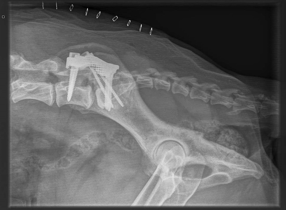
Image credit: RAM3D Ltd. Retrieved from https://www.ram3d.co.nz
Same species, model and patient
Total hip replacement is the clearest example of a technology where the line between "human medicine borrowing an animal model" and "veterinary medicine treating its own patients" essentially disappears.
Early prosthetic hip designs were evaluated directly in dogs: a Teflon hip prosthesis in dogs was reported as early as 1965, and a stainless-steel and vinylidene-fluoride-resin hip prosthesis was evaluated in dogs in 1971, work that ran in parallel with, and informed, human orthopedic implant design. By the early 1980s, canine total hip replacement had matured into a defined surgical technique in its own right: a study at Ohio State University's veterinary hospital documented 132 canine total hip replacements performed on clinical patients between 1976 and 1980, establishing the operative approach still recognizable in the procedure today. (Olmstead et al., 1981)
The data behind it
That procedure isn't a research artifact anymore; it's an established option for dogs with hip dysplasia or degenerative joint disease:
- A ten-year multi-clinic UK registry tracked 1,852 dogs that together underwent 2,375 hip replacement surgeries, 72 percent received a single hip, 28 percent received both. (Allaith et al., 2022)
- The same registry reported 185 deaths over that period, only 11 of which were due to complications directly related to the replacement itself. (Allaith et al., 2022)
- Independent case series report functional success rates in the 83–95 percent range. (Horberg et al., 2020)
Why it can't be modeled on a bench
The question orthopedic research needs answered isn't just "does the material tolerate contact with bone", it's whether a metal-and-polyethylene joint can absorb load bearing, torsional stress across a full gait cycle, thousands of times a day, for years, while living bone continues to remodel around it. That's a question about the interaction between a fixed material and a dynamic biological system over time, and no bench test yet replicates it.
The Regulatory Framework Veterinary Research Operates Under
Every study described above, whether run to benefit human patients, veterinary patients, or both, sits inside the same three-layer oversight structure in the US:
- USDA/APHIS enforces the Animal Welfare Act through unannounced facility inspections. It's worth being precise about scope here: the AWA's definition of "animal" specifically excludes birds, rats of the genus Rattus, and mice of the genus Mus bred for research use, a carve-out written into the 2002 Farm Bill after a long legislative fight. Dogs, cats, and non-human primates, the species most relevant to the case studies above, are squarely covered. (USDA, n.d.)
- AAALAC International is a voluntary but rigorously audited accreditation body that many veterinary teaching hospitals and research institutions hold themselves to beyond the legal minimum.
- Institutional Animal Care and Use Committees (IACUCs) must include a veterinarian and a community member from outside the institution by law, and must review and approve every protocol before a single animal is enrolled.
The 3Rs
Layered on top of that legal structure is the scientific framework researchers are trained to apply to every protocol: the 3Rs, first laid out by zoologist William Russell and microbiologist Rex Burch in their 1959 book The Principles of Humane Experimental Technique.
- Replacement — using methods that avoid animal use in research altogether.
- Reduction — using the minimum number of animals needed for reliable results.
- Refinement — minimizing pain, suffering, and distress for the animals that must still be used [16].
These aren't slogans, they're the operating standard behind the studies cited above, from the lead-implant infection tracking in research dogs to the hip-replacement registries following clinical patients for a decade.
For anyone training in or practicing veterinary medicine, this framework isn't background information about a separate field. It's the same committee structure, the same inspection regime, and the same 3Rs discipline that governs research conducted at veterinary teaching hospitals and colleges, including the clinical and surgical work happening in orthopedics, cardiology, and internal medicine departments right now.
Why This Matters for the Conversation With Pet Owners
The common mistake in public discussion of this topic is treating "animal research" as something that happens to animals for humans' benefit. The insulin protocol, the pacemaker lead, and the hip prosthesis tell a more complicated and, frankly, more interesting story: dogs have been model, test subject, and patient in the same research lineage, sometimes within the same decade.
Replacement alternatives, organ-on-a-chip systems, computational models, cell culture, are real, growing, and already reducing the number of animals needed for certain kinds of studies. But for questions that depend on a living, integrated circulatory, skeletal, or endocrine system responding over time, they aren't yet a full substitute, and the researchers closest to this work are also the ones pushing hardest to get there.
Understanding this history, and being able to explain it accurately, is part of practicing veterinary medicine responsibly and speaking to clients with credibility rather than talking points in either direction. The same standards of humane, regulated research that produced the insulin in the fridge and the pacemaker on the surgical tray are the standards veterinary institutions like IIVER hold their own clinical and teaching programs to.
References:
- Chakir, K. et al., cited in Canine Model of Pacing-Induced Heart Failure. PubMed. https://pubmed.ncbi.nlm.nih.gov/29987830/
- Anesthetic management and complications during transvenous pacemaker implantation in dogs. Frontiers in Veterinary Science. https://www.frontiersin.org/journals/veterinary-science/articles/10.3389/fvets.2026.1754437/full
- Artificial cardiac pacemaker placement in dogs with a cohort of myocarditis suspects... ScienceDirect. https://www.sciencedirect.com/science/article/abs/pii/S1760273418300717
- A retrospective review of 146 active and passive fixation bradycardia lead implantations in 74 dogs. PMC. https://www.ncbi.nlm.nih.gov/pmc/articles/PMC5870196/
- Frederick Banting, Charles Best, James Collip, and John Macleod. Science History Institute. https://www.sciencehistory.org/education/scientific-biographies/frederick-banting-charles-best-james-collip-and-john-macleod/
- The Prevalence of Pet Diabetes in the United States. Patterson Veterinary. https://www.pattersonvet.com/blog/the-prevalence-of-pet-diabetes-in-the-united-states
- Data Shows Growing Prevalence of Diabetes Among U.S. Pets. PR Newswire, 2015. https://www.prnewswire.com/news-releases/data-shows-growing-prevalence-of-diabetes-among-us-pets-300180613.html
- Olmstead, M.L., Hohn, R.B., Turner, T.M. Technique for Canine Total Hip Replacement. Veterinary Surgery, 1981. https://onlinelibrary.wiley.com/doi/10.1111/j.1532-950X.1981.tb00628.x
- Allaith, S. et al. Outcomes and complications reported from a multiuser canine hip replacement registry over a 10-year period. PMC. https://pmc.ncbi.nlm.nih.gov/articles/PMC10087566/
- Greater trochanter morphology and association with patient demographics, surgical factors, and post-operative stem position... PMC. https://www.ncbi.nlm.nih.gov/pmc/articles/PMC8864880/
- Animal Welfare Act. National Agricultural Library, USDA. https://www.nal.usda.gov/animal-health-and-welfare/animal-welfare-act