
Introduction
Fractures involving the distal diaphysis of the radius and ulna are common in canine patients, particularly following trauma such as vehicular accidents or falls. Stabilization using plate osteosynthesis offers reliable alignment and load-sharing fixation in the radius.This article details the surgical approach for managing such fractures using a combination of cranio-medial plating of the radius and normograde intramedullary pinning of the ulna.
Case presentation
A 6 year old indie dog was presented with clinical signs of forelimb lameness, localized pain and swelling were subjected to radiographic examination.
Two orthogonal views confirmed a complete, transverse distal diaphyseal fracture involving both the distal radius and ulna.
Routine hematology, serum biochemistry and pre-anesthetic evaluation were performed to ensure patient fitness for surgery.
Broad-spectrum prophylactic antibiotics (Ceftriazone @ 20 mg/kg I/V) were administered 30 minutes prior to surgery.
Anesthesia and Positioning
General anesthesia was induced using propofol (2 mg/kg I/V) and midazolam (0.1 mg/kg I/V) following premedication with tramadol (2 mg/kg I/M).
Anesthesia was maintained with isoflurane in oxygen via endotracheal intubation. The patient was placed in lateral recumbency, with the affected limb uppermost and supported on a sterile drape.
Radiographic Findings
Before Surgical Intervention

AP (Anterior posterior) view
After Surgical Intervention

AP (Anterior posterior) view

Lateral view
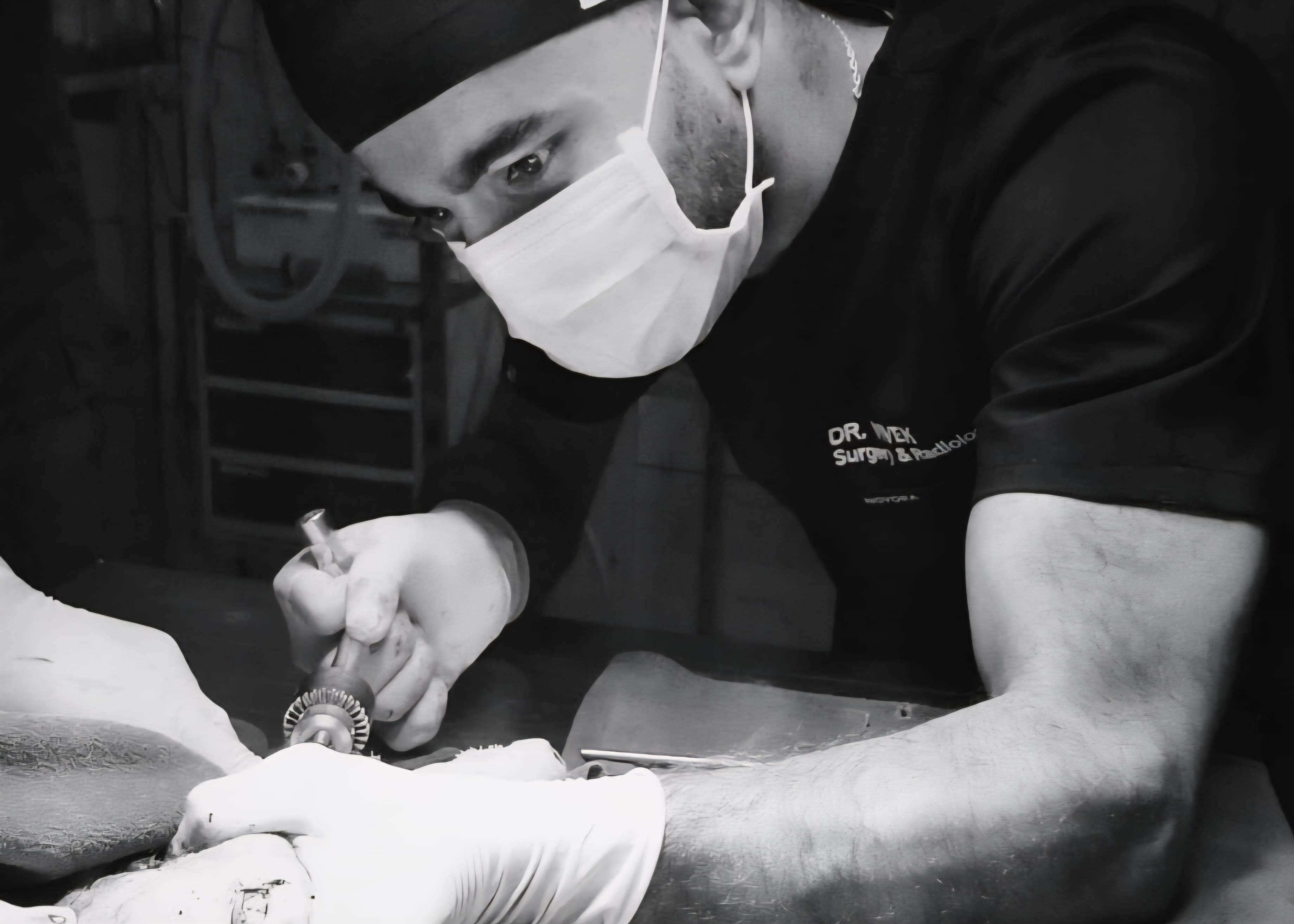
Surgical Technique
A cranio-medial approach to the distal radius was employed. A skin incision was made from the distal third of the antebrachium to the mid-diaphysis of the radius.
Subcutaneous tissues were bluntly dissected, taking care to preserve superficial veins and avoid injury to the cephalic vein. The extensor carpi radialis and abductor longus were gently separated to expose the cranio-medial surface of the radius.
Fracture Reduction and Plate Fixation
The fracture fragments were anatomically reduced using pointed reduction forceps.
A Limited contact dynamic compression plate (LCP) was selected based on the size of the patient and bone.
The plate was applied on the cranio-medial aspect of the radius and temporarily secured using bone-holding forceps.
Locking screws were placed on either side of the fracture using a standard drill–tap–screw technique.
Normograde Pinning of the Ulna
The proximal ulna was palpated just distal to the olecranon. A small stab incision was made at the proximal ulnar metaphysis.
A Steinmann pin of appropriate diameter (usually 2.0 mm) was introduced normogradely using a power drill.
The pin was directed distally into the medullary canal of the ulna, ensuring it did not exit the distal epiphysis or breach the carpal joint.
The periosteum and fascial layers were closed with 2-0 polyglactin 910 (Vicryl) in a simple continuous pattern.Subcutaneous tissue was apposed using 3-0 absorbable sutures.
Skin closure was completed with non-absorbable sutures (nylon 2-0) using an interrupted pattern.
Postoperative Management
- Postoperative analgesia was provided using carprofen (2.2 mg/kg PO BID) for 5 days.
- A modified Roberts jhones bandage was applied for 3weeks for Immobilization.
- Strict cage rest and activity restriction were advised for 6 weeks.
Outcome
The combination of radius plating and ulnar normograde pinning provided excellent stabilization, allowing for uneventful bone healing and return to limb function. No major postoperative complications such as implant failure, infection or delayed union were observed.
Conclusion
The described surgical approach offers a reliable, biomechanically sound and cost-effective method for managing distal diaphyseal radius-ulna fractures in dogs.
While the radius is stabilized with rigid internal fixation using a plate, the ulna can be effectively managed using a minimally invasive normograde pin.


.jpg)